
— A multinational team led by biodiversity researchers published a set of high-resolution three-dimensional X‑ray images of ants on Thursday, producing a public, searchable collection at antscan.info. Using a synchrotron accelerator in southwest Germany, the project scanned 2,200 preserved specimens drawn from museums and private collections during a concentrated, weeklong campaign. The images expose external and internal anatomy at submillimeter detail and are intended to accelerate research in taxonomy, ecology and comparative morphology. Project leaders say the dataset compresses work that would normally take years into a matter of days, creating a new resource for scientists and the public alike.
Key Takeaways
- The Antscan project produced 3‑D X‑ray scans of 2,200 ant specimens in a single intensive week at a synchrotron facility in southwest Germany.
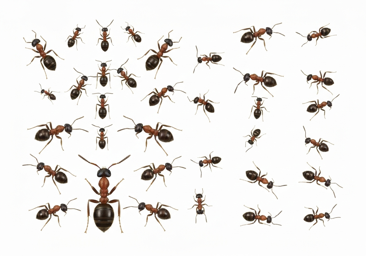
- Scans include species ranging from very large bullet ants (noted for potent stings) to microscopic specialists such as spider‑egg predators, representing global museum and private collections.
- The dataset and interactive renders are publicly available at antscan.info and were published alongside a methods paper in the journal Nature Methods.
- Approximately 20 quadrillion ants are estimated to exist on Earth, underscoring the project’s sampling relative to global abundance but highlighting remaining gaps in geographic and taxonomic coverage.
- The scanning run required round‑the‑clock shifts and specialized staff, including technicians at the Karlsruhe Institute of Technology.
- High‑throughput imaging and AI‑assisted pose reconstruction reduced time and labor compared with traditional micro‑CT workflows.
Background
Ants are among the most numerically abundant animals on Earth and play diverse ecological roles, from soil turnover and seed dispersal to predation. Their social complexity and wide range of body plans make them a focus for studies in behavior, evolution and ecosystem function. Yet detailed morphological data remain scattered across collections and publications, and many species lack modern digital vouchers that permit remote study.
Micro‑computed tomography (micro‑CT) has been used for years to visualize small organisms in three dimensions, but scanning thousands of specimens at museum‑grade resolution has typically been prohibitively slow and expensive. Synchrotron facilities, which produce highly focused X‑ray beams, can accelerate throughput; Antscan combined such imaging with automated processing to scale scans across hundreds of samples per day. The project partnered with museums and private collectors to assemble a geographically and taxonomically wide set of specimens for scanning.
Main Event
The scanning campaign took place over a single, concentrated week at a synchrotron facility in southwest Germany managed in part by staff from the Karlsruhe Institute of Technology (KIT). Teams worked in shifts around the clock to load specimens, collect raw X‑ray data and run reconstruction pipelines. Organizers say what would normally take years of staggered micro‑CT work was completed in days by taking advantage of the accelerator’s intense beam and optimized workflows.
Specimens were sourced from multiple institutions and private collections; organizers prioritized representation across ant subfamilies and body sizes. Technicians imaged large species such as Eciton and Paraponera alongside tiny specialists that feed on spider eggs and other narrowly targeted niches. After reconstruction, the team applied machine learning routines to render the poses more lifelike and to segment anatomical regions for downstream analysis.
Project leaders emphasized open access: the complete set of scans and 3‑D visualizations are hosted at antscan.info to enable researchers, educators and the public to inspect morphology without handling fragile specimens. The accompanying methods description was submitted to Nature Methods, documenting scanning parameters, reconstruction software and quality controls so other groups can replicate or extend the approach.
Analysis & Implications
By compressing a traditionally slow imaging workflow into a weeklong run, Antscan demonstrates how centralized facilities and pipeline automation can scale morphological digitization. This matters for taxonomy, where high‑quality morphological vouchers speed species descriptions and comparisons, and for phylogenetics, where form complements genetic data. The availability of standardized 3‑D models will reduce barriers for researchers who lack access to physical collections.
The dataset’s size and scope also have conservation implications: accurate morphology aids in identifying cryptic or undescribed species that may be at risk. Yet the scans alone do not substitute for field data; distribution, population trends and ecological interactions must still be gathered in situ. Antscan functions best as a complementary resource that amplifies museum holdings rather than replaces fieldwork.
Scaling this approach globally could reveal regional biases in collections and accelerate automated trait extraction, but it will require sustained coordination and funding. Synchrotron beamtime is limited and costly; broad adoption will depend on hybrid strategies that pair large‑facility runs with distributed micro‑CT at regional centers and improved sample pipelines to reduce handling time.
Comparison & Data
| Metric | Value |
|---|---|
| Specimens scanned | 2,200 |
| Estimated ants on Earth | ~20 quadrillion |
| Scanning campaign duration | 1 week (continuous shifts) |
| Typical small‑scale workflow | Months–years per comparable sample set |
The table highlights the project’s throughput versus conventional approaches. While 2,200 specimens is numerically small compared with global ant diversity, the uniform imaging standards and public availability multiply the dataset’s value for comparative studies. Future runs that prioritize underrepresented regions and taxonomic groups would make the resource more comprehensive.
Reactions & Quotes
The team running the synchrotron described the project as a rare demonstration of high‑throughput biodiversity imaging made possible by facility scale and coordination.
Thomas van de Kamp, Karlsruhe Institute of Technology (project technician)
Van de Kamp was part of the staff who operated the accelerator and noted the practical intensity of the run, including overnight shifts and rapid sample changes to maintain throughput.
The lead biodiversity scientist framed the scans as a bridge between traditional collections and modern digital science, emphasizing utility for taxonomy and ecosystem research.
Evan Economo, University of Maryland (biodiversity scientist)
Economo’s team coordinated specimen selection and algorithmic post‑processing; he highlighted the value of standardized, open 3‑D vouchers for both specialists and educators.
Unconfirmed
- Whether every scanned specimen has been taxonomically verified to current nomenclatural standards; some identifications may be pending expert revision.
- The extent to which scanning altered any specimen labels or mounting hardware during handling has not been fully documented in public materials.
- Long‑term plans for additional scanning campaigns, including specific funding commitments and timelines, were described in broad terms but not confirmed in formal public announcements.
Bottom Line
Antscan’s public release of 2,200 high‑resolution 3‑D ant scans represents a practical demonstration of how centralized imaging resources and automated pipelines can expand access to morphological data. The collection will be valuable for taxonomists, comparative biologists and educators who need reliable, manipulable digital vouchers without moving fragile specimens.
At the same time, the project is an early step rather than a comprehensive solution: broader geographic and taxonomic coverage, integration with field data and sustainable funding models are required to realize the full potential. For now, Antscan provides a scalable template and a usable dataset that other institutions can cite, replicate and build upon.
Sources
- New York Times — media report summarizing the Antscan project and interviews with researchers (journalism).
- Antscan — project website hosting the public 3‑D scans and interactive renderings (project/official resource).
- Karlsruhe Institute of Technology (KIT) — host institution for the synchrotron used in the scanning campaign (research facility/institutional).
- Nature Methods — journal where the methods paper was published (academic journal).