
Lead
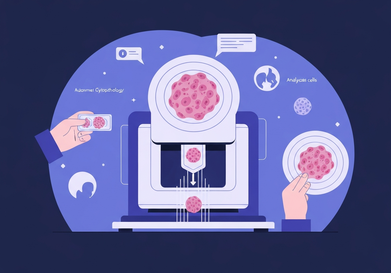
Researchers report a real‑time, autonomous cytology pipeline that combines high‑resolution three‑dimensional whole‑slide imaging with on‑device (edge) computing and AI to classify cervical cells with clinical‑grade performance. Published in Nature on 18 February 2026, the system digitizes thick liquid‑based cytology preparations (ThinPrep and SurePath) at 40 Z layers per slide and processes each slide in minutes, delivering population‑level cell counts that correlate with HPV status and cytological diagnoses. A single‑centre clinical test used 318 held‑out donor slides and a multicentre evaluation covered 1,124 slides across four centres, producing slide‑level AUCs near 0.9 for LSIL+ and HSIL+ detection. The authors present an interpretable population‑analysis embedding — the cluster of morphological differentiation (CMD) — to visualise and gate morphologic cell phenotypes at scale.
Key takeaways
- The imaging system captures high‑resolution bright‑field frames (4,480 × 4,504 pixels) at up to 50 fps and acquires 40 axial layers per slide, producing ~140 gigavoxels for SurePath and ~391 gigavoxels for ThinPrep slides.
- Optical resolution is 220 nm laterally and 1 μm axially; whole‑slide acquisition times were ~3 min (10 layers), ~4.5 min (20 layers) and ~8 min (40 layers) depending on Z depth settings.
- Edge processing using FPGA + SOM (Jetson Xavier NX) builds and HEVC‑compresses sectional 3D stacks on the device, reducing data transfer burdens while preserving diagnostic fidelity at PSNR ≥ 40 dB.
- Automated nucleus detection used a YOLOX‑based detector trained on 242,669 annotated nuclei (348 images); classification used a MaxViT model trained on ~168,569 augmented single‑cell images.
- Clinical evaluation: single‑centre test (318 slides) produced AUCs of 0.84 (LSIL+) and 0.89 (HSIL+); multicentre AUCs ranged ~0.86–0.97 across four sites (n = 1,124 slides).
- CMD encodes a 10–11 dimensional class‑probability vector per cell to enable scatter gating, histograms and UMAP visualisations for population‑scale interpretability and discovery of morphological trajectories.
- AI counts of LSIL/HSIL cells track with HPV positivity: in NILM slides, HPV+ cases had significantly higher AI‑detected abnormal cell counts (adjusted q values q = 0.005 for LSIL, q = 0.038 for HSIL in the single‑centre subset with HPV data).
Background
Cytology—especially liquid‑based Pap testing—remains a cornerstone of early detection for cervical and other epithelial cancers because it is minimally invasive, inexpensive and widely deployable. In routine practice, cytologists review slides containing from ~10,000 up to 1,000,000 cells, relying on 3D nuclear and cytoplasmic morphology and spatial relationships to make diagnostic judgements.
Despite its public‑health impact, visual cytology suffers from inter‑ and intra‑observer variability driven by training, cognitive biases, fatigue and workflow pressures; these limitations can produce missed or delayed diagnoses and contribute to high‑profile screening failures. Prior AI work has largely focused on 2D images of selected fields or representative cells, limiting applicability to whole‑slide workloads and to the three‑dimensional cues that human experts use.
Three‑dimensional imaging can capture richer morphological information but increases demands on acquisition speed, processing, storage and network transfer. The combination of volumetric cytology and scalable AI therefore requires innovations both in imaging hardware and in data handling strategies so whole slides can be digitized, compressed and analysed in clinically acceptable timescales.
Main event
The team implemented a whole‑slide edge tomograph that pairs a high‑resolution CMOS sensor (IMX531) and motorised XY/Z stages with an edge computer containing an FPGA and an SOM (NVIDIA Jetson Xavier NX). The camera captures 4,480 × 4,504 bright‑field frames at up to 50 fps and collects either 173 or 485 imaging sections per layer depending on preparation, with 40 depth layers per slide in the high‑resolution protocol.
On the edge unit the FPGA performs initial signal conditioning and the SOM executes background correction, focus selection, 3D stack assembly and HEVC hardware‑accelerated compression (NVENC). The pipeline exploits intra‑layer and inter‑layer redundancy (akin to video intra/inter‑frame prediction) to compress volumetric stacks into HEVC files that preserve diagnostic features while reducing file sizes (e.g., ~1 GB, 500–800 MB, or ~170 MB for a ten‑layer SurePath slide at high/medium/low quality).
Compressed sectional volumes are transmitted to back‑end servers for GPU‑accelerated stitching into full whole‑slide 3D volumes and for on‑demand tile decoding in a deep‑zoom viewer. The viewer supports fast random access (most tile requests completed <100 ms) and Z‑plane navigation so cytologists can inspect reconstructed volumes interactively while AI analysis runs in parallel.
AI processing begins with a high‑sensitivity YOLOX detector (trained on 242,669 nuclei) run on 3 μm subsampled Z stacks to find nuclear centroids. For each grouped nucleus the best‑focused Z slice is chosen and a 224 × 224 patch extracted; a MaxViT‑based classifier then outputs a 10–11 dimensional probability vector per cell. These vectors form the basis of the CMD population analysis used for gating, histograms and UMAP visualisations.
Analysis & implications
From a technical perspective, the combination of edge‑side compression and accelerated AI inference addresses a practical bottleneck: volumetric cytology produces very large data volumes, and transferring full raw stacks to a central server would be costly and slow. By performing reconstruction and HEVC compression at the edge, the system enables routine digitisation of thick cellular clusters while keeping latency low enough for near‑real‑time review.
Clinically, slide‑level quantitative counts of LSIL and HSIL cells provided discriminative power: single‑centre AUCs were 0.84 (LSIL+) and 0.89 (HSIL+), and multicentre AUCs averaged near 0.9. These figures indicate the pipeline can stratify lesion severity robustly across institutions and preparation methods (SurePath vs ThinPrep), suggesting utility both as an assistive prescreen and, in some settings, as a triage tool for HPV‑positive screening populations.
The CMD embedding is an important interpretability advance: rather than returning opaque labels, the system supplies per‑cell probability vectors that clinicians can explore with scatter plots, gating and UMAPs to verify AI detections, locate rare abnormal cells, and discover morphological continuums (for example, trajectories from parabasal to superficial squamous phenotypes). This supports auditability and discovery while reducing reliance on single‑cell manual picks.
However, several barriers remain before broad deployment: the datasets used for training and validation are substantial but proprietary (raw images are restricted for privacy and IP reasons), prospective outcome‑linked trials are needed to show improved patient outcomes, and regulatory approvals and workflows must be addressed to integrate autonomous outputs into diagnostic pathways safely.
Comparison & data
| Metric | Value |
|---|---|
| Lateral / Axial resolution | 220 nm / 1 μm |
| Frames / sensor | 4,480 × 4,504 pixels @ up to 50 fps |
| Voxels per slide | ~140 Gvox (SurePath), ~391 Gvox (ThinPrep) |
| Detector training | 242,669 nuclei (348 images) |
| Classifier training | ~168,569 augmented single‑cell images |
| Clinical cohorts | 318 test slides (single centre); 1,124 slides (multicentre) |
| Slide‑level AUCs | Single centre: 0.84 (LSIL+), 0.89 (HSIL+); Multicentre: 0.86–0.97 range |
The table summarises core hardware, dataset and diagnostic performance numbers reported by the authors. AUC stability was observed across a range of per‑cell confidence thresholds (0.60–0.90), and performance was robust across SurePath and ThinPrep preparations.
Reactions & quotes
“We built an integrated, real‑time pipeline that digitizes thick cytology samples and produces interpretable, population‑level morphology maps with clinical‑grade predictive performance,” the study authors write, summarising the platform’s goals and outcomes.
Authors (Nitta et al., Nature 2026)
“Edge‑side compression plus on‑device inference addresses a critical bottleneck for volumetric cytology: it makes whole‑slide 3D imaging operational in routine workflows without prohibitive data transfer costs,” the methods team notes in their technical description.
Study technical team
“CMD transforms single‑cell outputs into an exploratory framework — enabling gating, histograms and UMAPs — which helps reconcile AI outputs with human review and facilitates discovery of intermediate phenotypes,” the paper emphasises.
Study authors
Unconfirmed
- Long‑term clinical impact: whether implementation of the platform will reduce cancer incidence or mortality in population screening remains unproven and requires prospective trials with outcome endpoints.
- Regulatory and deployment timelines: the paper does not specify timelines for regulatory approvals or commercial roll‑out in different jurisdictions.
- Generalisability to other specimen types: while the authors show representative non‑cervical examples (breast, thyroid), broad validation across non‑gynaecological cytology domains is not yet reported at scale.
Bottom line
This work demonstrates that whole‑slide, volumetric cytology can be made clinically practical by integrating high‑speed optical tomography with edge computing and modern deep‑learning models. The platform achieves near‑clinical performance for slide‑level detection of low‑ and high‑grade cervical abnormalities while providing interpretable, population‑scale visualisations through the CMD framework.
If validated prospectively with outcome measures and integrated into regulatory‑compliant workflows, this approach could reduce subjectivity in cytology, improve triage for HPV‑positive screening, and expand access to high‑quality diagnostic services—especially where cytotechnologist capacity is limited. Remaining priorities are independent prospective trials, regulatory clearances, and transparent pathways for data sharing and external validation.
Sources
- Nature — peer‑reviewed article (Nitta et al., Clinical‑grade autonomous cytopathology, 18 Feb 2026)
- Zenodo — data & code release (anonymized CSVs and source code archive) (repository noted in the paper as public supporting materials)
- Cancer Institute Hospital of JFCR — clinical partner (institutional source)
- NVIDIA — hardware used (Jetson Xavier NX, NVENC/NVDEC) (vendor technical reference)