
Lead
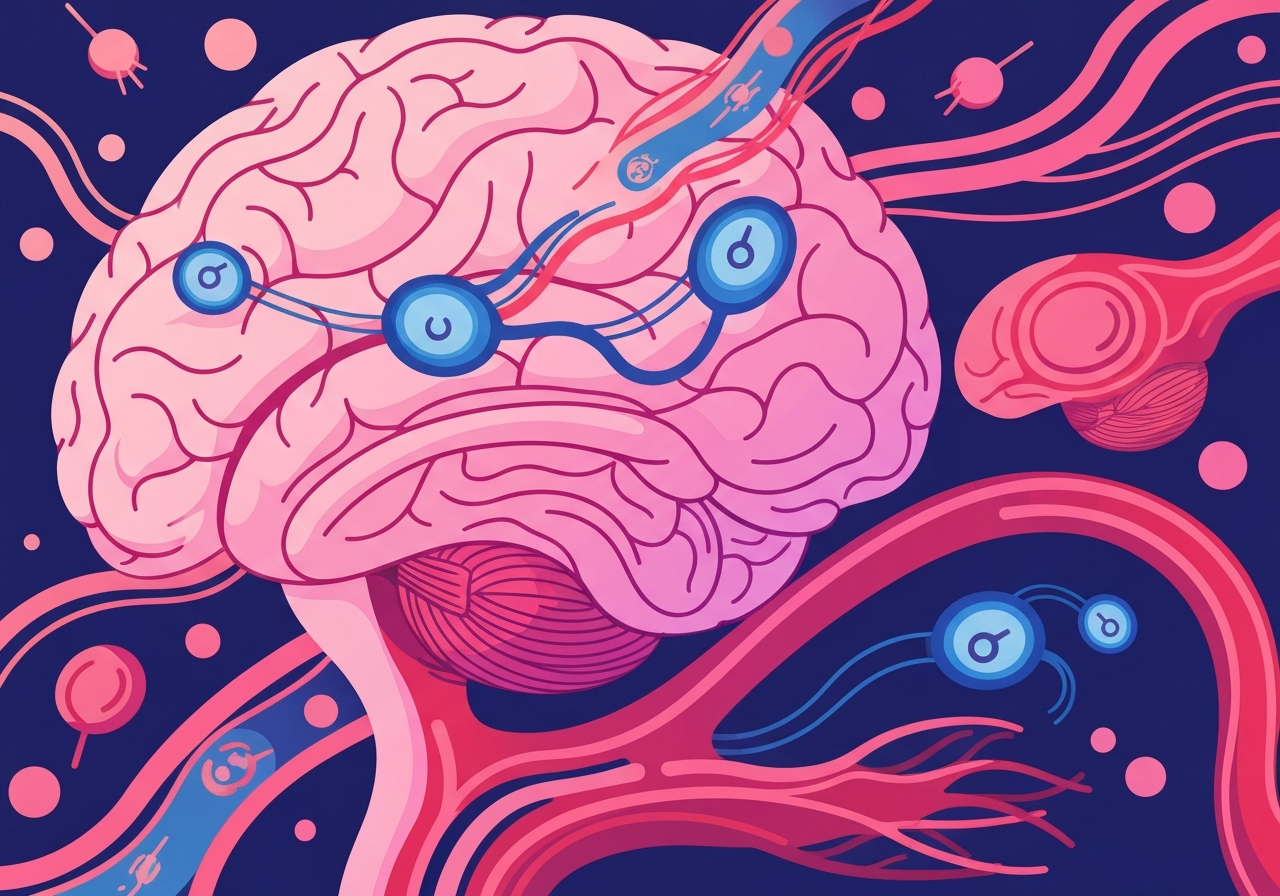
A recent study published in Nature reports that tanycytes — specialized cells lining the brain’s third ventricle — actively transport tau protein out of the cerebrospinal fluid (CSF) into the bloodstream. Researchers led by Vincent Prévot (Inserm, Paris) found this clearance route in cell models and mice, then identified disrupted tanycyte function and fragmented tanycytes in people with Alzheimer’s disease. In human samples (86 people with Alzheimer’s, 91 controls) the team observed reduced movement of p-tau181 from CSF into blood in the Alzheimer’s group, implicating tanycyte failure in pathological tau accumulation. The work suggests a previously underappreciated clearance pathway that could affect diagnosis and therapy development.
Key takeaways
- Tanycytes line the third ventricle and connect CSF with the bloodstream, enabling bidirectional molecular transport, according to the new study.
- In cell cultures and in vivo mouse experiments with fluorescently labeled human tau, researchers observed tau uptake by tanycytes and delivery to the pituitary and then circulation.
- Mice with experimentally impaired tanycytes retained more tau in the brain and showed lower tau levels in blood, indicating a clearance deficit when tanycytes are dysfunctional.
- Human cohort analysis compared 86 people diagnosed with Alzheimer’s to 91 controls and found relatively less p-tau181 in blood versus CSF among the Alzheimer’s group.
- Postmortem tissue from people with Alzheimer’s showed fragmented or degraded tanycyte structures compared with controls, suggesting structural loss associated with disease.
- Findings point to a potential mechanism for tau accumulation and raise implications for blood-based tau biomarkers and therapies aimed at enhancing clearance.
Background
Tanycytes are a distinct class of ependymal-like glial cells that form a cellular interface along the floor of the third ventricle. Unlike most brain cells separated by the blood–brain barrier, tanycytes contact both CSF and blood vessels, positioning them to ferry molecules across compartments. Tau is a microtubule-associated protein that stabilizes neuronal cytoskeleton; in Alzheimer’s disease tau becomes abnormally phosphorylated (for example p-tau181), aggregates into intracellular tangles, and correlates with neurodegeneration and memory loss.
Clearance of misfolded proteins from the brain involves multiple routes: proteasomal and lysosomal degradation, glymphatic flow, and transport across barriers into peripheral circulation. Prior research emphasized endothelial and lymphatic routes, but tanycytes have been less studied in neurodegenerative contexts. Because blood-based tau assays are now used clinically and in trials, understanding cellular pathways that move tau into blood is critical for interpreting biomarker levels and for designing interventions that enhance protein removal.
Main event
The authors first demonstrated tau uptake and release in cultured tanycyte-like cells, showing those cells internalize tau and secrete it on the vascular-facing side. To test the route in vivo, they injected fluorescently tagged human tau into the CSF of mice and mapped its distribution over time. Fluorescent tau was detected predominantly within tanycytes and later in the pituitary region and systemic circulation but not broadly across other ependymal populations.
When researchers impaired tanycyte function experimentally, mice accumulated more tau within brain tissue and had reduced tau levels detectable in blood, supporting a causal role for tanycytes in clearing CSF tau into the bloodstream. The team then analyzed paired CSF and blood samples from 86 people with Alzheimer’s and 91 people without dementia; they observed proportionally lower p-tau181 in blood relative to CSF in the Alzheimer’s group, consistent with reduced transport.
Finally, postmortem histology revealed structural disruption of tanycytes in brains from people with Alzheimer’s disease. The cells appeared fragmented or degraded compared with control brains, a morphological finding the authors interpret as consistent with impaired clearance capacity in patients. Taken together, the experiments across models and human samples build a chain of evidence linking tanycyte dysfunction to reduced tau export from the central nervous system.
Analysis & implications
If tanycytes contribute substantially to removing tau from the CSF, their dysfunction could help explain how tau accumulates inside neurons and spreads across brain regions in Alzheimer’s disease. That offers a mechanistic complement to hypotheses focused on intracellular proteostasis failure and glymphatic decline. Importantly, the direction of causality remains to be established: tanycyte damage could be an upstream driver of tau pathology or a downstream effect of disease processes.
Clinically, the findings affect interpretation of blood-based tau biomarkers. Reduced transfer of p-tau181 into blood could mean that blood levels underestimate brain pathology in patients with tanycyte loss, potentially complicating screening and longitudinal monitoring strategies that rely on peripheral measures. Conversely, therapies that restore or enhance tanycyte transport might increase peripheral tau clearance and improve clinical outcomes, but could also change biomarker trajectories in unexpected ways.
Therapeutic targeting of tanycytes raises both opportunity and challenge. Because these cells sit at a neuroendocrine interface, modulating them could impact hormonal regulation and other homeostatic systems; safety will be a central concern. The result opens avenues for research into molecular drivers of tanycyte fragility, whether metabolic, inflammatory, or age-related processes prime these cells for degeneration, and whether interventions can preserve or restore their structure and function.
Comparison & data
| Group | Number of individuals | CSF p-tau181 | Blood p-tau181 | Interpretation |
|---|---|---|---|---|
| Alzheimer’s | 86 | Elevated | Relatively lower vs CSF | Suggests impaired transfer from CSF to blood |
| Controls (no dementia) | 91 | Lower | Relatively higher vs CSF | More efficient CSF-to-blood movement observed |
The table summarizes cohort sizes and qualitative biomarker patterns reported by the study. The authors did not publish absolute concentration values in the press summary; instead the comparison emphasizes relative distribution between compartments. Inter-individual variability, timing of sample collection and disease stage will influence exact measurements, so follow-up studies with standardized protocols and larger cohorts are needed to quantify effect sizes and diagnostic thresholds.
Reactions & quotes
Experts not involved in the research highlighted the novelty of implicating tanycytes in Alzheimer’s-related clearance. They noted that the study broadens the set of cellular players considered in protein homeostasis and raises testable hypotheses for how peripheral biomarkers reflect central pathology.
“Tanycytes are highways for the brain.”
Vincent Prévot, Inserm (lead author)
Prévot used the metaphor to emphasize the anatomical role of tanycytes as conduits between CSF and blood. The paper documents both transport activity in experimental models and structural alteration in human disease, linking the anatomical observation to potential functional consequences.
“No one has looked at these cells before in relation to Alzheimer’s disease.”
Amy Brodtmann, Monash University (commenting neurologist)
Brodtmann’s reaction underscores that tanycytes have been understudied in neurodegeneration. She and others called for additional human-focused work to clarify when tanycyte dysfunction arises in the disease timeline and whether it correlates with cognitive decline.
Unconfirmed
- Whether tanycyte breakdown is a primary driver of tau accumulation or a consequence of upstream Alzheimer’s pathology remains unresolved and requires longitudinal human studies.
- It is unconfirmed whether restoring tanycyte function would meaningfully reduce brain tau loads or improve cognitive outcomes in patients.
- The exact molecular triggers that fragment tanycytes in Alzheimer’s brains — whether inflammatory, metabolic, age-related, or tau-mediated — are not yet established.
Bottom line
The study identifies tanycytes as a plausible cellular pathway for moving tau from the CSF into blood and shows that tanycyte disruption correlates with reduced tau transfer and increased brain retention in experimental systems and human tissue. This reframes part of Alzheimer’s pathology as not only a problem of intracellular aggregation but also of impaired intercompartmental clearance.
For clinicians and researchers, the findings encourage caution when interpreting blood-based tau levels and motivate new lines of investigation into tanycyte health as a biomarker and therapeutic target. Next steps should prioritize longitudinal human sampling, quantification of clearance rates across disease stages, and mechanistic work to test whether preserving or augmenting tanycyte transport changes disease trajectories.
Sources
- Nature (Peer‑reviewed journal article / news report)
- Inserm (French National Institute of Health and Medical Research — research institute)
- Monash University (Academic institution / expert affiliation)