
Northwestern University surgeons kept a 33-year-old man alive for 48 hours after removing both lungs, using a bespoke extracorporeal circuit while they treated a life‑threatening infection and awaited donor organs. The patient arrived with Influenza B complicated by a carbapenem‑resistant Pseudomonas aeruginosa infection that caused necrotizing acute respiratory distress syndrome (ARDS), leaving his lung tissue irreversibly destroyed. Faced with refractory septic shock and failing organs, the team performed a bilateral pneumonectomy and connected the patient to a custom “total artificial lung” system that managed gas exchange and circulatory loading long enough for a successful double‑lung transplant. Two years after the procedures the report says he has returned to independent life with excellent lung function.
Key Takeaways
- The patient was 33 years old and presented with Influenza B and a secondary, carbapenem‑resistant Pseudomonas aeruginosa infection that produced necrotizing ARDS.
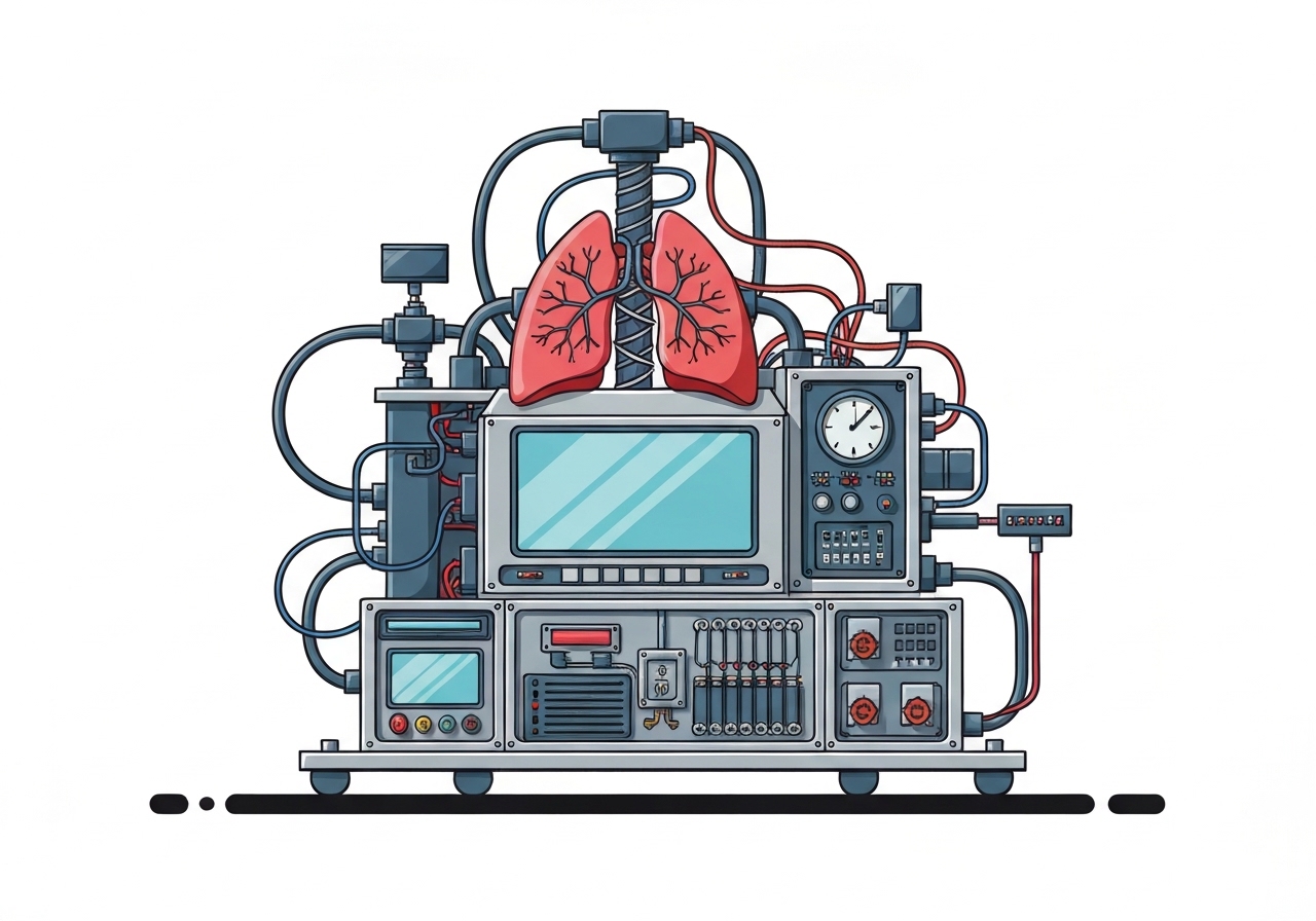
- Surgeons performed a bilateral pneumonectomy and supported the patient for 48 hours using a flow‑adaptive extracorporeal total artificial lung (TAL) developed at Northwestern.
- The TAL combined a pump/oxygenator with four novel elements: a dual‑lumen drainage cannula, a flow‑adaptive pulmonary shunt (1.1–6.3 L/min), dual left‑atrial return grafts (two 10 mm grafts), and mechanical chest reconstruction.
- Key physiological improvements occurred rapidly: lactate fell from 8.2 mmol/L to <1.0 mmol/L within 24 hours, and vasopressor infusions were stopped after about 12 hours.
- The patient was bridged for 48 hours until suitable donor lungs were available, underwent successful double‑lung transplantation, and was reported well at two‑year follow‑up.
- The clinical data and spatial transcriptomics of the removed lungs showed near‑complete loss of regenerative stem cells and replacement by aberrant basaloid and scar‑forming cells, defining an irreversible injury pattern.
Background
ARDS is a syndromic end stage of severe lung injury in which inflammation and fluid accumulation prevent adequate oxygen transfer into the blood. In many ARDS cases clinicians continue maximal support in the expectation that injured lungs will recover, and lung transplantation is rare for acute infectious ARDS because patients are frequently too unstable to survive surgery. Necrotizing infections, however, can progress to a point where lung parenchyma loses its capacity for repair—what the authors describe as a clinical point of no return.
Removing both lungs (bilateral pneumonectomy) is ordinarily avoided because the pulmonary vascular bed normally buffers the right heart: without it the right ventricle faces catastrophic pressure overload, and systemic circulation collapses unless circulation and preload are carefully managed. Standard ECMO provides gas exchange but does not replicate the pulmonary circuit’s mechanical role; in the empty thorax ECMO use is associated with major bleeding, heart malposition, clotting, and stroke risks. The Northwestern team therefore designed an integrated mechanical solution to restore both oxygenation and the missing hemodynamic load while treating sepsis.
Main Event
The patient’s lung tissue was liquefying from a necrotizing infection and was judged unlikely to recover. With kidneys failing and a cardiac arrest early in his admission that required resuscitation, clinicians elected to remove both lungs to eliminate the source of persistent sepsis. Performing a bilateral pneumonectomy under these conditions is exceptionally high risk, so the team prepared an extracorporeal circuit to replace key pulmonary functions immediately after explant.
The TAL architecture combined an oxygenator and pump from standard ECMO with four bespoke parts to manage right‑ and left‑sided circulation. A dual‑lumen cannula positioned via the internal jugular vein drained deoxygenated blood from the right heart, actively unloading the right ventricle. A flow‑adaptive shunt linked the right pulmonary artery to the right atrium; that shunt automatically diverted excess right‑sided output when the extracorporeal pump reached its limit, with measured flows between 1.1 and 6.3 liters per minute during support.
To preserve left‑sided filling and Starling physiology, oxygenated blood returned via two 10 mm grafts directly into the left atrium (dual left atrial return), preventing chamber stagnation and reducing intracardiac clot risk. Surgeons rebuilt the pericardial envelope using bovine pericardium and filled the mediastinal space with tissue expanders and sponges to stabilize the heart’s position and protect bleeders while the chest remained lungless.
The physiological impact was rapid: septic shock reversed, lactate normalized within 24 hours, and vasopressors were discontinued after roughly 12 hours. The team kept the patient on TAL support for 48 hours while controlling infection and arranging donor lungs; once suitable organs arrived, the patient underwent successful double‑lung transplantation and later recovered sufficient function for normal independent living at two years.
Analysis & Implications
This single‑case intervention demonstrates that, with tailored engineering and surgical steps, it is possible to temporarily replace both the gas exchange and the mechanical vascular role of the lungs and bridge a patient through otherwise lethal sepsis to transplant. The dual approach—controlling both right‑sided unloading and left‑atrial filling—addresses the core physiological challenge created by removing the pulmonary vascular capacitance. That combination appears central to preventing acute right‑ventricular failure and systemic collapse.
The molecular mapping of the explanted lungs provides a complementary diagnostic advance: spatial transcriptomics revealed a depletion of regenerative stem cells and a dominance of aberrant basaloid and fibrotic cell populations, offering objective markers of irreversibility. If these tissue signatures are validated in larger cohorts, they could help identify which acute patients will not recover with supportive care alone and therefore should be considered for early transplant evaluation.
However, the approach has limitations that constrain immediate generalization. It requires high‑level cardiac and transplant surgery expertise, specialized perfusion equipment and personnel, and rapid access to donor lungs—resources concentrated in few centers. The procedural risks—bleeding, stroke, infection of the extracorporeal circuit and technical failure—remain significant and must be weighed against potential benefit on a patient‑by‑patient basis.
Looking ahead, the TAL concept could inspire modular extracorporeal strategies that extend bridge times and broaden transplant candidacy, but adoption will depend on reproducible outcomes, multicenter protocols, and careful patient selection. Regulatory and logistical issues around wider use of paired surgical‑engineering solutions will also need resolution before dissemination beyond specialized academic centers.
Comparison & Data
| Parameter | Normal lungs intact | TAL support during pneumonectomy |
|---|---|---|
| Right‑sided buffer (pulmonary capacitance) | Present: physiologic vascular bed | Replaced: flow‑adaptive shunt 1.1–6.3 L/min |
| Gas exchange | Alveolar diffusion | ECMO oxygenator within TAL |
| Left atrial preload | Maintained physiologically | Dual left‑atrial return via two 10 mm grafts |
| Clinical bridge time achieved | Not applicable | 48 hours (successful bridge to transplant) |
The table summarizes how TAL substituted both mechanical and gas‑exchange roles of native lungs in this case. TAL’s measured flow range and direct left‑atrial return aimed to restore preload and protect against right‑ventricular overload—two functions standard ECMO alone cannot fully reproduce when lungs are absent.
Reactions & Quotes
Lead surgeon Ankit Bharat and colleagues framed the procedure as a deliberate decision to remove the infection source and reconstruct the missing circulatory functions mechanically. They argue the molecular data from the removed lungs help delineate when injured lungs are beyond salvage and when transplantation should be considered.
“When the infection is so severe that the lungs are melting, they’re irrecoverably damaged.”
Ankit Bharat, Northwestern University (surgical lead)
This statement was used by the team to justify the operative choice to explant rather than continue expectant support. The authors emphasize that in some young patients with fulminant necrotizing infection, waiting for recovery can be futile and that earlier transplant consideration may save lives when irreversible tissue loss is documented.
“In my practice, young patients die almost every week because no one realized that transplantation was an option.”
Ankit Bharat, Northwestern University (surgical lead)
Bharat’s comment underscores a clinical gap: acute transplant pathways for rapidly deteriorating patients are uncommon, and the team’s work aims to expand those pathways. The report is careful to note, however, that the approach requires exceptional institutional capability and that conclusions are drawn from a single, carefully selected case.
Unconfirmed
- Whether the TAL configuration and tissue markers of irreversibility will be reproducible across different hospitals and patient populations remains unproven; the report describes a single case.
- The applicability of the observed spatial transcriptomic pattern (aberrant basaloid cells and stem‑cell depletion) to ARDS from other pathogens or stages of disease is not yet validated.
- Longer‑term complication rates attributable specifically to this approach (stroke, late thrombosis, graft infection) require larger cohorts and systematic follow‑up.
Bottom Line
This case demonstrates that a custom extracorporeal system can temporarily replace both the gas‑exchange and mechanical vascular functions of the lungs, allowing surgeons to remove a deadly source of infection and bridge a patient to transplantation. Objective tissue mapping from the explanted lungs gives clinicians a molecular framework to identify irreversible lung injury, a potential tool to refine transplant candidacy in acute settings where time is critical.
Wider use of the TAL concept will hinge on multicenter validation, standardized selection criteria, and the resources to perform such complex care. For now, the report offers a carefully documented proof of principle: in exceptional centers, engineering and surgical teams can successfully rescue patients once thought unsalvageable, but broader impact will depend on reproducibility and access.